
Micropatterned Cell Models Reveal How Geometry Controls Endoplasmic Reticulum Organization During Epithelial Wound Closure
Understanding how epithelial tissues respond to geometric constraints provides important insights into the mechanisms that regulate collective cell migration during wound closure. A recent study published in Nature Cell Biology explores the relationship between epithelial edge curvature and intracellular organization, revealing that the morphology of the endoplasmic reticulum (ER) is dynamically reorganized in response to wound-edge curvature and correlates with distinct modes of epithelial migration.
To investigate these mechanisms, researchers cultured epithelial cells on micropatterned substrates engineered to reproduce concave and convex wound-edge geometries. These controlled experimental models enabled the analysis of how local curvature influences ER organization and cellular migration behaviour during wound closure.
Precise Micropatterning Enables Controlled Cell Biology Studies
The micropatterned substrates used throughout the study were fabricated using a Durham Magneto Optics (DMO) MicroWriter direct-write lithography system, enabling the production of well-defined micrometric patterns that reproduced specific epithelial wound geometries for quantitative analysis under different curvature conditions.
By providing controlled geometric environments, micropatterned substrates allow researchers to dissect how physical parameters, such as edge curvature, influence cellular behaviour and intracellular organization. In this study, this approach was essential for comparing epithelial responses at concave and convex wound edges and for examining the relationship between tissue geometry, cytoskeletal forces, and organelle organization.
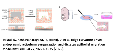
Figure 1. A schematic representing the experimental set-up for creating gaps of definite geometries. Cells surround the PDMS stencils placed on glass and after their removal the cells migrate into the voids.
Edge Curvature Directs Endoplasmic Reticulum Reorganization
Using these micropatterned models, the researchers showed that the endoplasmic reticulum undergoes curvature-dependent reorganization during epithelial migration. At convex wound edges, the ER predominantly adopted tubular structures, while at concave wound edges it reorganized into sheet-like structures.
The study demonstrated that these changes are associated with cytoskeleton-generated protrusive and contractile forces, highlighting the connection between cellular mechanics and ER architecture. The researchers also found that different ER organizations were associated with distinct focal adhesion orientations and corresponded to two modes of epithelial migration: lamellipodial crawling at convex regions and purse-string contraction at concave regions.
Together with mathematical modelling, these findings identify the endoplasmic reticulum as a key mechanotransducer that integrates geometric and mechanical cues during collective epithelial migration.
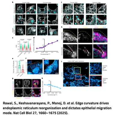
Figure 2. b, Representative images of actin, microtubule and ER at convex (left) and concave (right) edges. MDCK cells stained with phalloidin (grey, actin marker) and DAPI (cyan) (top), anti-α-tubulin (grey, microtubule marker) and DAPI (cyan) (middle), and MDCK cells expressing mApple-Sec61β (grey, ER marker) and labelled with DRAQ5 (cyan) (bottom). The edge of the cells near the curvature is enlarged on the right of individual images. Scale bar, 10 μm. c, MDE quantifications for microtubules, actin and ER at convex (green) and concave edges (pink). From left to right: n = 62, 69, 74, 62, 84 and 76. d, The fraction of ER tubules at the front as a function of curvature, n = 50 at convex and n = 72 at concave. e, Quantification of the fraction of ER tubules present at the front of the cells at the convex (green) and concave (pink) edges. From left to right: n = 50 and 52. f, Representative super-resolution images of MDCK cells expressing mApple-Sec61β at convex (left) and concave (right) edges. Insets: tubules at the front (left) and sheets at the front (right). g, Representative images of cells stained with anti-Climp63 (yellow, ER sheet marker), phalloidin (pink) and DAPI (cyan) at the convex (top) and concave (bottom) edges. The blue arrowheads show low Climp63 at the edge of the cell and the white arrowhead shows Climp63 enrichment at the edge. Scale bar, 10 μm. h, Quantification of the fraction of Climp63 at the front of the cell edge as a function of curvature, n = 74 at convex and n = 104 at concave.
High-Precision Lithography for Advanced Cell Biology Research
This study highlights the importance of precisely engineered micropatterned substrates for investigating how physical geometry influences cellular organization and behaviour. The DMO MicroWriter direct-write lithography system enables researchers to fabricate defined micrometric patterns, providing a controlled approach for creating cellular microenvironments to study cell migration, mechanobiology, and the relationship between cellular structures and physical constraints.
For further technical details about the DMO MicroWriter lithography system and its applications in advanced research workflows, contact our expert Marwan Channab or read the product page.