
Electron Diffraction: the missing link between SC-XRD and XRPD
Single-crystal X-ray diffraction (SC-XRD) provides the most complete structural information available, including unit-cell parameters, space group and atomic positions. However, it requires crystals typically tens to hundreds of micrometres in size.
At the other end of the scale, X-ray powder diffraction (XRPD) is ideally suited to bulk powders and remains a key technique for identifying dominant crystalline phases. Yet its one-dimensional diffraction patterns can make structure solution challenging, particularly when peaks overlap or minority phases are present. Phases below approximately 1–5% by mass may not be reliably detected.
This creates a significant analytical gap for nanocrystalline materials, samples with suspected trace polymorphism, or materials for which XRPD provides ambiguous results.
3D Electron Diffraction: structural resolution from powder samples
3D Electron Diffraction (3D-ED) provides a complementary approach between SC-XRD and XRPD.
Electrons interact with matter roughly 1,000 times more strongly than X-rays of comparable energy. In a 3D-ED experiment, a focused electron beam is directed onto an individual crystal within a standard powder sample, typically 50–1000 nm thick, while the sample is continuously rotated. Diffraction data collected from multiple orientations can provide the same fundamental structural information obtained by SC-XRD: unit-cell parameters, space group and atomic positions.
The crucial advantage is therefore the ability to obtain crystal-structure information from crystals that are too small for conventional SC-XRD.
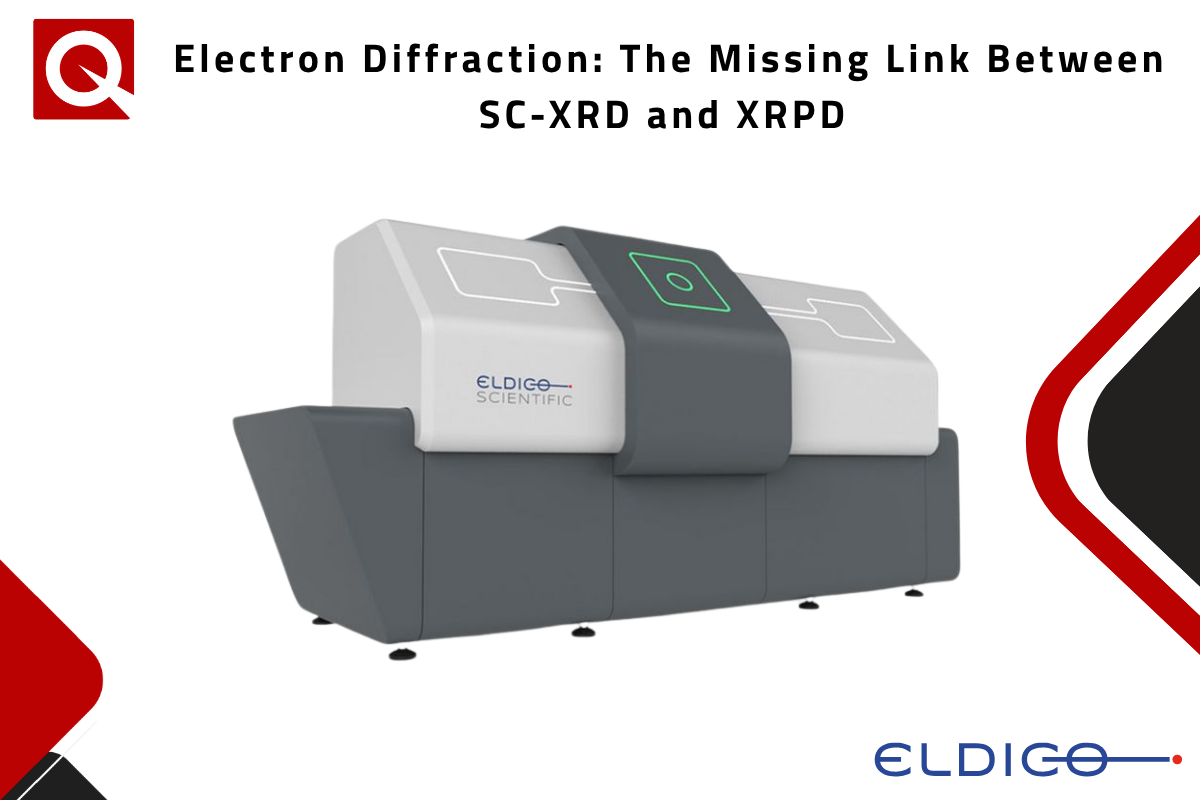
Developed by ELDICO Scientific, the ELDICO ED-1 combines dedicated instrumentation with automated data collection and crystal mapping. These capabilities enable systematic screening of individual crystals within powder samples and facilitate the detection and characterisation of different crystalline phases.
Key Applications of 3D Electron Diffraction
For nanocrystalline APIs, it can enable structure determination directly from powder material when crystal growth to X-ray-compatible dimensions is not feasible. In the case of dabigatran etexilate mesylate, two anhydrous forms and a hemihydrate were characterised from nanocrystalline powder, supporting solid-form selection.
3D-ED is also particularly valuable for polymorph identification and minority phases. In an automated study of commercial dapsone, approximately 650 crystals were surveyed in 24 hours; around 3% showed an unknown unit cell, leading to the identification of a previously unknown polymorphic form, Dapsone Form VI.
Finally, 3D-ED can reveal residual crystallinity in materials appearing amorphous by XRPD. In carvedilol samples prepared by melt quenching and ball milling, electron-diffraction crystal mapping showed the melt-quenched material to be fully amorphous, while the ball-milled sample retained crystallinity.
A complementary diffraction workflow
3D-ED does not replace SC-XRD or XRPD Instead, the three techniques provide complementary information:
- SC-XRD: complete structural information from sufficiently large single crystals.
- XRPD: bulk phase characterisation of powders.
- 3D-ED: structural information from individual crystals within powder and nanocrystalline samples.
Explore ELDICO’s 3D Electron Diffraction solution
Contact our expertMarwan Channabto discuss how 3D electron diffraction could support your research needs.
| +39 06 5004204 | |
| +39 06 5010389 | |
| marwan.channabqd-europe.com |