New measuring technique proves exceptional temperature accuracy of Wildfire Nanochip

by Lama Elboreini
The temperature-sensitive luminescence of nanoparticles enables their application as remote thermometers. In fact, the size of these nanothermometers makes them ideal to map temperatures with a high spatial resolution. Yet, conducting high spatial resolution mapping of temperatures that exceed 100°C poses some challenges.
In collaboration with Thomas van Swieten and his fellow colleagues at Utrecht University, DENSsolution was able to jointly develop a new technique to measure temperature at the nanoscale. In fact, they tested this novel technique on our Wildfire Nanochip and were able to further confirm the Nanochip’s unparalleled temperature accuracy and homogeneity. These experiments also proved how well their models work to predict the temperature distribution across the microheater. Importantly, this particular technique will improve the accuracy of nanothermometry as a whole, not only in micro- and nano-electronics but also in other fields with photonically inhomogeneous substrates.
The technique: Luminescence nanothermometry
Thermometry on the microscopic scale is an essential characterization tool for the development of nano- and microelectronic devices. However, conventional thermometers like thermocouples are often unable to reliably measure the temperature on this length scale due to their size. This is precisely where remote temperature sensing via optical thermometry techniques comes into play. Thermometry based on luminescence is particularly interesting since it is easily implemented, requiring only the deposition of a luminescent material in or on a sample of interest and the detection of its luminescence. For this reason, luminescence nanothermometry is currently developing into the method of choice for temperature measurements in microscopy.
Homogeneous heat distribution
DENSsolution's Wildfire Nanochip was specifically designed to enable users a homogeneous heat distribution across the microheater where a sample is positioned. It is particularly due to the unique geometry of the metal spiral, where the windows are placed right at the center, that users are able to enjoy such a remarkable temperature homogeneity. In fact, this Wildfire Nanochip has a temperature uniformity of 98% across the window area and 99.5% across the two central windows. The figure on the right below is a perfect illustration of the chip’s exceptional temperature homogeneity, showing the temperature profile across the membrane and the microheater for a center temperature of 523 K simulated with a finite element model.
The high temperature homogeneity of the Wildfire Nanochips is also owed to the fact that the metal heating spiral is embedded in a silicon nitride membrane. Silicon nitride has many advantages including being chemically inert, mechanically robust and can withstand harsh chemical and temperature environments.
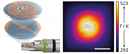
On the left: The Wildfire Nanochip, where the metal spiral is represented in orange and the silicon nitride membrane in blue.
On the right: Finite element model simulation showing the remarkable temperature homogeneity of the Wildfire Nanochip
Reliable temperature mapping
In this work, the luminescent particles that were used are NaYF₄ nanoparticles doped with Er³⁺ and Yb³⁺. These particles exhibit a strong upconversion when excited with an infrared laser. In other words, they emit photons with a shorter wavelength than the excitation photons. As shown in the figure below, they found that the spectrum of the emitted (green) photons is quite sensitive to temperature.
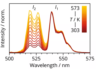
Green upconversion luminescence of the nanoparticles upon excitation at various temperatures ranging from 303 K (dark red) to 573 K (yellow)
By scanning the laser across a layer of deposited nanoparticles in the confocal microscope, they were able to capture an array of emission spectra. They then converted this emission spectra into a temperature map using the luminescence intensity ratio of the 2 peaks at each pixel. After a number of correction steps, the technique showed a remarkable precision of 1-4 K with a spatial resolution of ∼1 micrometer. It is noteworthy to mention that most other techniques are unable to achieve such a high accuracy like this.
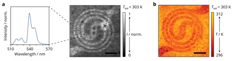
In a) they scanned the laser across the microheater with the deposited luminescent nanoparticles to generate a map of intensity ratios.
b) shows the spectrum at each pixel converted into a temperature to provide a temperature map of the microheater.
Simulation and model accuracy
Using the fully corrected temperature maps, they were able to analyze in depth the temperature homogeneity of the microheater. The figure below shows the horizontal traces through the center of these maps. The simulated temperature profiles (lines) show an excellent match with the experimental traces (dots).This confirms both the reliability of the finite element model as a design tool and the strength of our temperature mapping technique as a characterization tool, achieving a high accuracy and a spatial resolution of ∼1 μm.
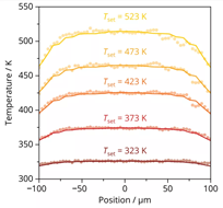
A graph showing the mapping of elevated temperatures. The lines represent the simulated temperature profiles and the experimental traces represent the dots.
They determine the standard deviation of the temperature in the center to quantify the accuracy of this thermometry method and find values of 1 K at 323 K increasing to only 4 K at 513 K. Conclusively, this makes nanothermometry using confocal luminescence spectroscopy a promising method to map temperature profiles not only for microheaters but also in other fields such as biology and catalysis where temperature variations are important but hard to monitor with conventional methods.
Click here for more info on Wildfire in situ TEM heating series
