The Prisma XRM – a new class in x-ray 3D-tomography
The Prisma XRM from Sigray defines a new class in the field of X-ray tomography and X-ray microscopy. The founder of Sigray, Mr. Wenbing Yun as already proven with his first company Xradia that there are new possibilities in the field of X-ray tomography. Xradia established the field of so-called X-ray microscopes (XRM) in the laboratories around the world which breaks with the traditional plat panel detector design that was used and which is still used in commercial available laboratory X-ray ct-systems. This traditional design is using a nanofocus X-ray source and a flat panel detector behind the sample and the reachable resolution is driven by the geometrical magnification of the system. Primarily by the size of the focus of the X-ray source, the distance between source to sample and the distance sample to detector. This means that you need to bring your sample as close to the source as possible to get the best possible resolution.
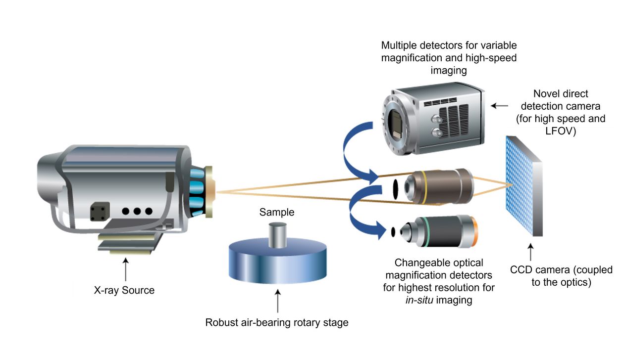
Mr. Wenbing Yun thought about a different approach to overcome these limitations and to achieve an even better resolution. He implemented a new detector approach by inserting microscope objectives with a very thin scintillator film on top. These objectives had different magnifications and this whole new X-ray system was in principle comparable to a traditional optical microscope. That is the reason why this new class of systems is called X-ray microscope instead of the traditional name Computer Tomography systems (or µCT system). The general internal setup of such an XRM can be seen in figure 1 and it has the following major advantages:
The overall resolution of the system is no longer not only depending on the size of the focus spot of the X-ray source and the source to sample distance but has now a second magnification step with the used optical objective.
This means that we are much more independent from the geometrical magnification, and we can now use much larger source to sample distances which will open the road to in-situ experiments while keeping the achievable resolution still as high as possible.
Wenbing Yun was now able to get to resolutions beyond 500 nm while increasing the possibilities in the field of in-situ X-ray experiments.
The new Sigray Prisma XRM system is now pushing the limits even further. It is using not only one of the best nanofocus X-ray sources that is available in the market but can in addition equipped with a second innovative X-ray source developed by Sigray. The first nanofocus source can be used for a variety of different samples while the second source is primarily focusing on softer samples made out of low z materials (e.g. life science or polymer science samples). These softer samples are very hard to measure with traditional-ray sources as those sources are using most often target materials like Tungsten which simply generate too much X-ray energy (which is very good for hard materials) but which is not the best solution for such delicate and light sample materials. Therefor Sigray developed the new Chroma source (please see Spectrum E37) and implemented it into the new Prisma XRM system to cover both types of samples, hard as well as soft materials.
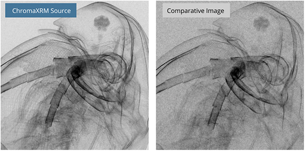
Figure 2 is showing the impact that the Target material can have to the image quality. You see a Life Science sample measured with the Chroma source on the left (target material was Chromium) and the same sample measured with a traditional Tungsten target. You clearly can see that the Chroma source produces a much sharper image with more internal details compared to the traditional Tungsten source. Another highlight of the Chroma x-ray source is the possibility to equip this source with up to 5x different target materials. This will increase the capabilities of the source even further.
This makes the Prisma XRM to the most powerful XRM and CT-system up to date. In combines the requirements that a multi-user facility has into one cutting edge system. You have the best possible resolution (<500 nm) and the flexibility of different sources and different target materials to measure all type of samples. No matter if these samples are from Material Science departments or from the Life Science departments.
We are open to measure your samples to prove that this system is really the most powerful system that one can find.


