SCREEN Cell3iMagers for robust and high-throughput profiling of 3D microtissues growth and morphology

3D microtissues or spheroids are more and more used in drug discovery and development as a more representative biological model for testing drug sensitivity and efficacy. Studies using spheroids and organoids are increasingly important in phenotypic drug discovery recently, due to their biological relevance. Size and morphology are important determinants to evaluate the biological behavior of 3D spheroids, particularly in the development of anti-cancer drugs where monitoring cell growth is a critical, translatable endpoint. The ability to monitor spheroid growth in terms of size provides a similar unit of measure with which to make direct in vitro to in vivo comparisons.
The increasing number of applications of 3D spheroids as an in vitro model for drug discovery requires their adaptation to large-scale screening formats in every step of a drug screen, including high-throughput image analysis. Although the benefits of 3D cell culture models have been widely acknowledged, widespread implementation of spheroids in drug discovery screening efforts has been hindered by lack of appropriate companion assays and instrumentation.
Historically, the process of imaging spheroids to monitor growth using conventional or even automated microscopes has been slow and tedious. High-throughput, accurate assessment of 3D microtissue size and growth in these applications is currently limited by sequential, time-consuming and often manual optical measurements. SCREEN designed innovative instrumentation (Cell3iMagers) and software solutions to overcome such kind of speed and throughput limitations in phenotypic screening.


Select the instrument which best reaches the imaging needs for your cell type
| Cell3iMager duos (cc-8000) | Cell3iMager neo (cc-3000) | Cell3iMager (cc-5000) | |
|---|---|---|---|
| Well plates | 6,12, 24, 48, 96 and 384 wells, flat and U-shaped, 35 mm dish, T-5 flasks | 6,12, 24, 48, 96 and 384 wells, flat and U-shaped, 35 mm dish, T-5 flasks | 6,12, 24, 48, 96 and 384 wells, flat and U-shaped, 35 mm dish, T-5 flasks |
| Number of plates | 1 | 1 | 4 |
| Scanning speed (96 well plate) | 90 s/plate | 50 s/plate | 54 s/plate |
| Scanning resolution | 0.8 µm & 4.0 µm/pixel | 2.6, 5.0 & 10.0 µm/pixel | 2.6, 5.0 & 10.0 µm/pixel |
| Light mode | Bright field & fluorescent | Bright field only | Bright field only |
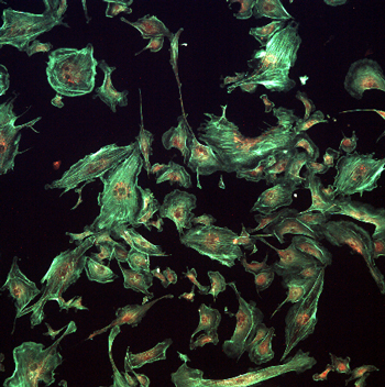
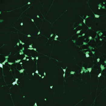
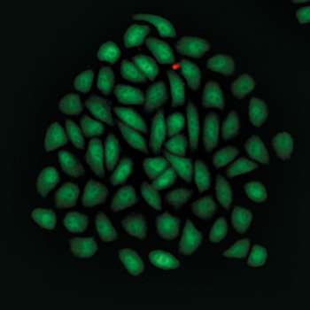
| Measurements | Single cells, dead/living cells, number of cells, total number, spheroid area, pseudo volume, circularity and diameter of spheroids, neurite length etc. | Number, area, pseudo volume, circularity, optical density and spheroid diameter | Number, area, pseudo volume, circularity, optical density and spheroid diameter |
| Focus | Auto focus, manually, multi-level Tumor cells and cell lines Primary cells and co-cultures | Auto focus, manually, multi-level Tumor cells and cell lines Primary cells and co-cultures | Auto focus, manually, multi-level Tumor cells and cell lines Primary cells and co-cultures |
| Image quality | +++ | ++ | ++ |
These instruments are useful in many research settings, including phenotypic drug discovery, drug sensitivity testing, combinatorial drug testing, and drug-target discovery and validation, co-culture associated loss of spheroid volume, spheroid quality control, and many more.
Key features
- Escape throughput and speed limitations in phenotypic assessment of 3D microtissue size and morphology (scans a 384-well plate in <1 minute)
- Only cost effective, non-invasive solutions for kinetic growth profiling of 3D microtissues
- Bright-field and fluorescence imaging systems capable of scanning one to four plates depending on the instrument type
- Determination of spheroid number, size, and growth kinetics, time dependent analysis of rate of cell proliferation, aggregate count, monoclonality detection, neurite length measurement
- Analysis of single/multi-spheroids per well; images spheroids, organoids, iPSc, monolayer culture etc.
- Comprehensive 3D image analysis package with built-in compatibility for all major 3D cell culture platforms


