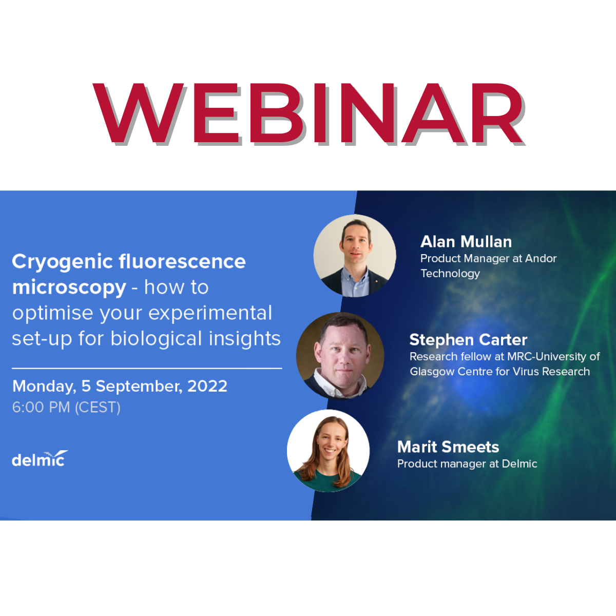
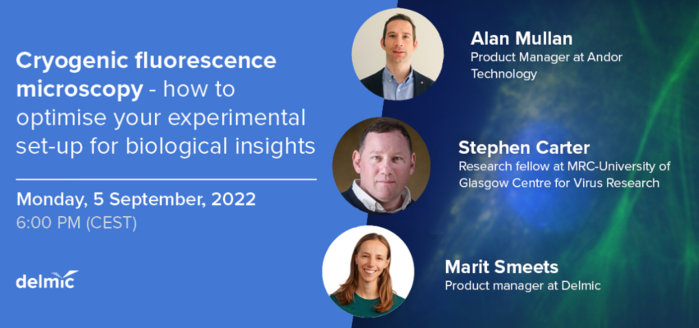
Webinar: Cryogenic fluorescence microscopy – how to optimise your experimental set-up for biological insights
Monday, 5th September, 2022 - 6:00 pm CEST
Cryo-FLM is often used to identify the region of interest to prepare lamella for cryo-ET since it increases the success rate in capturing the structure of interest in the lamella.
The benefits of cryo-FLM in the cryo-ET workflow are undoubted but its usefulness can be compromised if the optical design of the system is not optimised for detecting the weaker fluorescence signals from the lamella or does not allow the artefacts inherent to fluorescence microscopy to be overcome.
In this webinar, the Cryo Product Manager Marit Smeets from our partner company Delmic will be joined by Dr Stephen Carter, Group Leader at MRC-University of Glasgow Centre for Virus Research, and Dr Alan Mullan, Product Manager for microscopy cameras at our partner company Andor Technology.
Together, the expert speakers will walk you through the crucial aspects of cryo-FLM imaging to help you get more reliable cryo-FLM data.
Join them to get answers to your cryo-FLM-related questions!
The webinar will see three talks, as follow:
- Avoiding excitation crosstalk in multi-channel fluorescence imaging - Ms Marit Smeets
- Subtracting autofluorescence inherent to cryo-FLM - Dr Stephen Carter
- Choosing a scientific camera for cryo fluorescence microscopy - Dr Alan Mullan, Product Manager @ Andor Technologies
The recording of the webinar will be available right after the end for all registrants.
CLICK HERE TO REGISTER!
Below you can find the Abstracts of the three talks:
Avoiding excitation crosstalk in multi-channel fluorescence imaging - Ms Marit Smeets
Cryogenic fluorescence microscopy (cryo-FLM) combined with scanning electron microscopy (SEM) makes the process of lamella milling in the cryo-electron tomography (cryo-ET) workflow more accurate and efficient. The benefit of cryo-FLM can however be compromised if the optical design of the system does not allow the users to easily overcome the artifacts inherent to fluorescence microscopy.
Excitation crosstalk is an artifact that can appear when two or more fluorescent labels with partially overlapping excitation and emission spectra are used to tag different types of biomacromolecules. In this presentation, we will discuss the cause of excitation crosstalk and how this can be prevented by selecting the correct optical components.
Subtracting autofluorescence inherent to cryo-FLM - Dr Stephen Carter
Cryogenic correlated light and electron tomography (cryo-CLEM/cryo-ET) is a powerful technique to study the complex relation between protein complexes and organelles inside cells. Typically, cryo-CLEM uses fluorescent proteins to initially locate a protein-of-interest inside large eukaryotic cells using cryo-fluorescence microscopy. This fluorescence information allows coordinates of a protein-of-interest to be mapped for precise localization of the same target inside the electron microscope and subsequent high-resolution structural interrogation using cryo-ET. A major challenge for researchers that can hamper the identification of the “real” fluorescent protein signal in the cryo-fluorescence microscope is the presence of bright “autofluorescence” at 80K.
In this talk, Stephen Carter will focus on how autofluorescence can be problematic for cryo-CLEM. He will provide practical examples detailing sources of autofluorescence in mammalian cells that have hindered his cryo-CLEM experiments and simple ways the autofluorescence problem can be overcome.
Choosing a scientific camera for cryo fluorescence microscopy - Dr Alan Mullan
Imaging cameras come in a wide range of different sensor formats and offer combinations of sensitivity, speed, and field of view. Furthermore, the same sensor may feature in a number of different cameras models, but the sensor can be implemented differently. These differences may appear to be quite subtle and thus unclear if they have any meaningful impact on imaging performance.
One difference between camera designs is how the sensor is cooled, and additionally how the sensor is enclosed. It is relatively well known that cooling the sensor reduces thermal noise (dark current) and this is important for applications like luminescence that require longer exposures into many minutes. But how valid is sensor cooling to the shorter exposures of typical fluorescence-based imaging that are measured in milliseconds, and how much cooling is needed?
In this presentation, we will discuss the topic of sensor cooling of sCMOS and CCD (and EMCCD) based cameras. We will look at how sensors may be cooled efficiently in terms of the overall camera design. As part of this, approaches to how the sensor is enclosed in the sensor chamber will be outlined. Then we will look at what parameters of the sensor are impacted by operating temperature and how this impacts sensor behaviour. From this we can determine if, and when we need to consider cooling for a given imaging application.


