


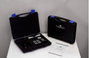
Correlative imaging
Correlative imaging solution from CorrescopyCorrelative imaging refers to the correlation of the data (usually in form of the images) collected from the single sample by different imaging techniques. This conventionally requires using correlative microscopes, however, Correscopy allows using any available imaging instrument such as microscope or spectrometer (spectroscope) to do so. Correlative imaging technique relies on the ability to correlate the information from a particular area of interest of the sample (usually in size of few to tens of µm2).
Imaging systems used to provide correlative imaging are usually very complex and expensive instruments. If the interest of correlative imaging arises, the asking price of the system is a major obstacle for the vast majority of users.
- Ease of use
- Independent - apply on any imaging instrument without it´s modification
- Versatile - works with almost every imaging techniques
- Affordable
Further information
Some of the laboratories build their own correlative solutions, which allow them to combine some of the imaging techniques for which they have an interest at the moment. However, this is rare, difficult to deliver and expensive. Correlative microscopes manufacturers, offer very limited choice of the techniques that can be correlated. It is caused, by the company-specific expertise in one or two imaging techniques. Moreover, the correlative systems, with have inbuild few imaging instruments, often involve a compromise on performance and data quality compared to the dedicated instruments.
Correscopy meets the demands of modern imaging by proposing a patent pending system which allows performing correlative imaging on any imaging instrument. Thanks to this, data correlation possibilities become almost unlimited. The solution does not require any modification of the instrument, it is an independent accessory that does not affect instrument’s standard operation. The obtained data can be easily compared and correlated using any data analysis software already available on the market. Correscopy provides undisputed data for making clear-cut conclusions.
Applications
Correlative imaging using Raman spectroscopy (Horiba LabRam HR) and scanning electron microscopy (Carl Zeiss Merlin)
In this experiment, a surface of an inhomogeneously corroded metal chip was examined. The Correscopy solution was used to correlate variations of Raman spectra against the surface features observed under a scanning electron microscope as a function of the location on the chip.

Correlative imaging using light microscopy (CLSM Carl Zeiss LSM 880) and atomic force microscopy (Bruker Catalyst)
In this experiment fibroblasts (cells) were imaged by two high-resolution techniques: light microscopy (Carl Zeiss LSM 880 with Airyscan) and atomic force microscopy (Bruker catalyst). The high-resolution fluorescence data about the cell structure is directly compared with the AFM results which provide e.g. the information about the mechanical properties of the living cell.Downloads


Contact

Navigation
Categories
Contact
Quantum Design SAS
Avenue de l’Atlantique
Bâtiment Fuji Yama
91940 Les Ulis
France
| Phone: | +33 1 69 19 49 49 |
| E-mail: | franceqd-europe.com |
