Cailabs beam shaping technologies for optical microscopy
Used for centuries for imaging and medical diagnosis, optical microscopes produce images of samples or objects that are invisible to the naked eye. Essentially consisting of a light source, lenses, mirrors, and an objective that magnifies the object to make it visible to the eye or camera, they generate images of living organisms or tissues on a cellular scale.
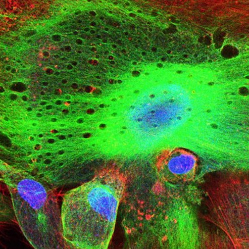
Different optical methods can be used to generate image contrast at the sample level: phase contrast, differential interference contrast and fluorescence excitation contrast. Fluorescence imaging is the most commonly used among optical microscopy techniques. It involves activating fluorescence by projecting an excitation laser beam of a certain wavelength on a fluorophore-marked sample using organic dyes, fluorochrome biomarkers, fluorescent proteins, etc. The fluorescence signal obtained is then used to visualize the organization of particles within the cells.
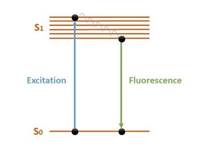
Schematic diagram of the fluorescence phenomenon
Optical microscopes can be divided into two main categories according to the sample analysis technique used:
- Widefield microscopy – the entire sample is exposed to a light source and the image is directly observable by the eye or a camera. This technique is ideal for viewing thin specimens perpendicular to the optical axis. This category includes both bright field and dark field microscopy.
- Laser scanning microscopy – a 2D or 3D image of the sample is generated using optical sectioning. This technique involves scanning the sample point by point with the laser beam. An algorithm is used to reconstruct the image, which is transmitted to the observer. Confocal microscopy and two-photon excitation microscopy use this technology to generate higher resolution images.
Widefield microscopy – the entire sample is exposed to a light source and the image is directly observable by the eye or a camera. This technique is ideal for viewing thin specimens perpendicular to the optical axis. This category includes both bright field and dark field microscopy.
Laser scanning microscopy – a 2D or 3D image of the sample is generated using optical sectioning. This technique involves scanning the sample point by point with the laser beam. An algorithm is used to reconstruct the image, which is transmitted to the observer. Confocal microscopy and two-photon excitation microscopy use this technology to generate higher resolution images.
- Stimulated Emission Depletion (STED) – this technique uses laser scanning and fluorescence microscopy to reduce the beam size by overlaying it with a donut-shaped depletion beam;
- Structured Illumination Microscopy (SIM) – the sample is illuminated with a structured light pattern (Moiré pattern) from different angles, at least nine angles for a 2D image. An algorithm is then used to reconstruct the image;
- Single-Molecule Localization Microscopy (SMLM) – this fluorescence microscopy method relies on the stochastic “blinking” of fluorophores phenomena to provide optimal spatial resolutions (STORM, d-STORM, PAINT, PALM).
However, microscopy devices still face some challenges. Cailabs’ customizedsystems improve the performance of optical microscopes:
- Gaussian laser beams are currently used in microscopes, and their profile is for the uniform imaging of samples. MPLC technology enables ultra-stable homogeneous beam shaping with an efficient use of laser energy, thus increasing process yield and quality. To improve fluorescence signal collection, a custom “top-hat” beam shape (e.g. square for Widefield microscopy, line for efficient laser scanning, ring, complex shape, etc.) adapts the illumination to the target area to obtain better quality images.(1)
- At reception – Cailabs proposes solutions to increase signal sensitivity and image resolution using a modal approach. Using MPLC as a mode analyzer exploits both the intensity and phase at the microscope output.(2)
Discover our Laser Beam Shaping Solutions here >>
References:
(1) Rowlands, C.J., Ströhl, F., Ramirez, P.P.V. et al. Flat-Field Super-Resolution Localization Microscopy with a Low-Cost Refractive Beam-Shaping Element. Sci Rep 8, 5630 (2018).
(2) Martin Poinsinet de Sivry-Houle, Simon Bolduc Beaudoin, Simon Brais-Brunet, Mathieu Dehaes, Nicolas Godbout, and Caroline Boudoux, “All-fiber few-mode optical coherence tomography using a modally-specific photonic lantern,” Biomed. Opt. Express 12, 5704-5719 (2021).
