Fast and versatile – The new SCREEN Duos2 opens up new possibilities for high throughput screening
Recent advances in imaging technology have resulted in better understanding of cellular processes and implications in the field of life sciences. Imaging technology has been widely used for characterizing cellular structures, assessing morphological and intercellular changes to understand the biological processes. Additionally, imaging technology has proven to be of immense advantage in high content and high throughput screening for drug discovery applications. In recent years, cell-based assays have been markedly improved to make in vitro assays physiologically relevant platforms to study and delineate changes at morphological as well as cellular levels. Therefore, 3D cell culture systems are now being widely used for in vitro and ex vivo studies not only restricted to drug discovery and investigative toxicology, but also used to study changes both at phenotypic as well as molecular levels, thereby making the platform useful for multiple disease therapeutic areas as well as in regenerative medicine.
The new SCREEN imaging technology platform Cell3imager DUOS 2 is capable of imaging both 3D as well as 2D cell cultures. The fluorescence capability of DUOS 2 platform allows to carry out multiparametric assays (2D and 3D) to observe changes at morphological, cellular, and molecular levels cultured in time lapse mode. This technology is capable of assessing and quantifying neurite outgrowth assays, non-invasive monitoring of spheroids and Organoids in addition to multifluorescence staining for providing quantitative information on protein/biomarkers of interest. The unparalleled fast bright field imaging capability of the system allows for non-invasive cell-based assays.
The new SCREEN imaging technology platform Cell3imager DUOS 2 is capable of imaging both 3D as well as 2D cell cultures. The fluorescence capability of DUOS 2 platform allows to carry out multiparametric assays (2D and 3D) to observe changes at morphological, cellular, and molecular levels cultured in time lapse mode. This technology is capable of assessing and quantifying neurite outgrowth assays, non-invasive monitoring of spheroids and Organoids in addition to multifluorescence staining for providing quantitative information on protein/biomarkers of interest. The unparalleled fast bright field imaging capability of the system allows for non-invasive cell-based assays.
The system comes with high resolution and high-speed scanning features with the possibility of integration within complete Robotics handling systems.
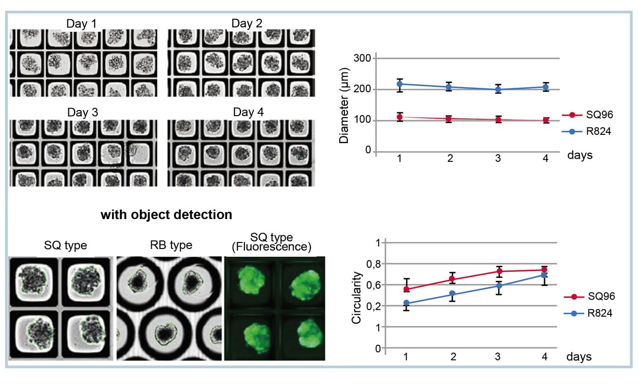
In the following two application examples the versatility of the system is highlighted. In the first one, intestinal organoids were cultured in microtiter plates and monitored for growth using label free bright field imaging. At the end of the culture period the organoid growth was extrapolated using Screen´s novel deep learning software technology as shown in figure 1.
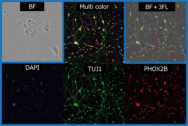
Emerging cell and molecular technology have resulted in utility of stem cells for studying the pathogenesis of various diseases. In this context, induced pluripotent stem cell (iPS cell) technology has provided an unique opportunity in regenerative medicine as well in neurodegenerative therapeutic area to study the cellular aspects of diseases using in vitro assay platforms for assessing compounds using neurite outgrowth end point measurements. As shown in figure 2, IPSC cells were cultured over a period of time and monitored for neurite outgrowth. The neurite lengths can be quantified to measure the effect of compounds. The neuronal cells were stained with TUJ1 (Tubulin beta III) and PHOX 2B to quantitate the cell bodies and neurite lengths.
In summary the Cell 3imager DUOS 2´s technology is robust, fast, and versatile for a broad spectrum of applications in cell-based assays for regenerative medicine and multiple disease therapeutic areas.


