Large area profilometry for topographic Raman imaging

TrueSurface Microscopy from WITec

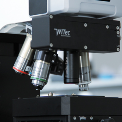
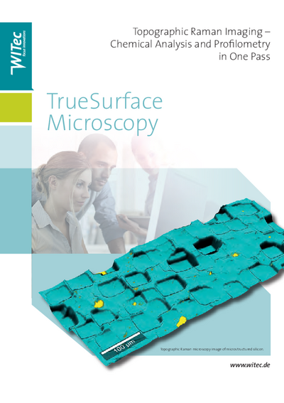
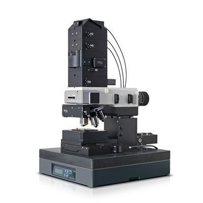
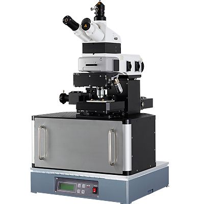
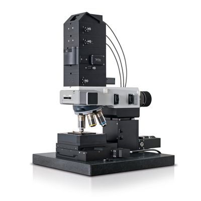
WITec´s award winning TrueSurface Microscopy allows confocal Raman imaging along heavily inclined or very rough samples with the surface held in constant focus while maintaining the highest confocality. The core element of this revolutionary imaging mode is an integrated sensor for optical profilometry. It is directly installed at the microscope turret which facilitates user-friendly and convenient handling.

Features
  • Unique combination delivers innovative application possibilities for new research techniques
  • Scan speed up to 2000 pixels/s for rapid data acquisition
  • Spatial resolution of 10 - 25 μm laterally and < 100 nm vertically to reveal an expanse of miniscule surface structures
  • Measuring distance: 10 mm - 16 mm providing wide-ranging sample size flexibility
  • Multi-sensors easily configurable to meet virtually any application

Further information

The TrueSurface Principle
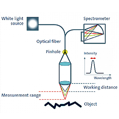
The key element of this novel imaging mode is a topographic sensor that works using the principle of chromatic aberration. With this non-contact, purely optical profilometer technique it is possible to trace a sample's topography and follow it in a subsequent Raman measurement, thus remaining in focus throughout.
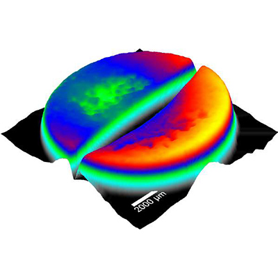
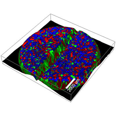
For profilometry a white light point-source is focused onto the sample with a hyperchromatic lens assembly: A lens system with a good point mapping capability, but a strong linear chromatic error. Every color has therefore a different focal distance. The light reflected from the sample is collected with the lens and focused through a pinhole into a spectrometer. As only one color is in focus at the sample surface, only this light can pass through the confocal pinhole. The detected wavelength is therefore related to the surface topography. Scanning the sample in the XY plane reveals a topographic map of the sample. This map can then be followed in a subsequent Raman image so that the Raman laser is always kept in focus with the sample surface (or at any distance below the surface). The results are images revealing chemical and/or optical properties at the surface of the sample, even if the surface is rough or inclined.

Applications

Life science From measurements in liquids to solid samples or soft tissues in life science varied samples are analyzed on a regular basis. With their convenient handling and versatile analytical capabilities the flexible WITec imaging systems provide the opportunity to adjust the imaging technique to changing requirements and are particularly well-suited for life science.
Pharmaceutics, cosmetics The development and production of drug delivery systems requires efficient and reliable control mechanisms to ensure the quality of the final products. These products can vary widely in composition and application. Therefore analytical tools such as the WITec imaging systems that provide both comprehensive chemical characterization and the flexibility to adjust the method to the investigated specimen are preferred in pharmaceutical research.
Materials science Materials science is a diverse field including the development and testing of new substances, as well as the refinement of manufacturing processes and quality control for existing products. WITec imaging systems are particularly well-suited for comprehensive sample analyses in materials science and provide the opportunity to acquire a thorough knowledge of the sample surface morphology and chemical composition.
Geoscience WITec confocal Raman imaging systems are excellent analytical tools for the comprehensive investigation of geological samples, such as the identification and characterization of minerals, or in the observation of mineral phase transitions in high and ultra-high pressure/temperature experiments.
Polymers WITec imaging systems enable comprehensive sample analysis that provides a thorough characterization of the physical and chemical properties of the polymers on the nanometer scale.
Nano-Carbon and graphene Nano-carbon materials such as graphene or carbon nano-tubes show immense promise in many applications such as transistors, sensors, and optoelectronics. Flexible and adaptive analytical methods can support effective investigation and accelerate progress in nano-carbon & graphene research and development.

Downloads

TrueSurface microscopy brochure
Chemical Characterization of Pharmaceutical Samples by Confocal Raman Microscopy and Correlative Techniques - AppNote
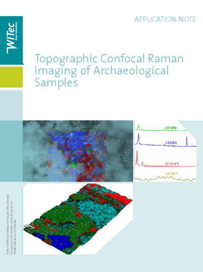
Topographic Confocal Raman Imaging of Archaeological Samples - AppNote

Contact

Stephane Struyve
Stephane Struyve

Related products

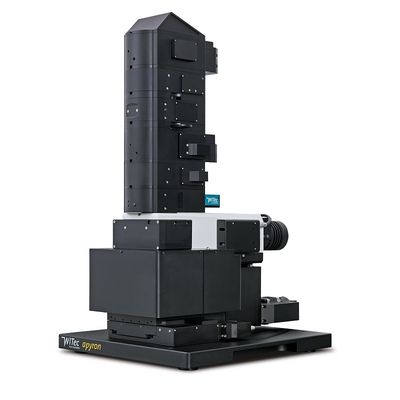
Automated 3D Raman imaging system
The alpha300 apyron is the top of the line Raman imaging system in WITec’s microscope series. It combines ease-of-use and ultimate capability by automating hardware control and offering pre-configured ...
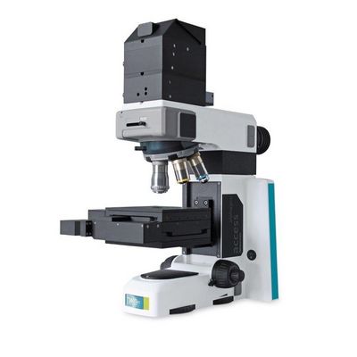
Basic Raman system
The alpha300 Access is a high-quality yet affordable micro-Raman system that provides the same outstanding performance and spectral resolution that other WITec products are renowned for. This unique ...
Chemical and nanoscale imaging system
The well-established Raman-atomic force microscope (AFM) combination alpha300 RA was the first integrated Raman AFM system on the market and continues to set the standard for combined instrument ...
Raman, AFM and SNOM all-in-one system
The unprecedented all-in-one alpha300 RAS combines Raman, AFM, and SNOM imaging in a single instrument for the utmost flexibility and sophisticated sample characterization. By combining the imaging ...
Superior confocal Raman imaging system
The imaging capabilities of the alpha300 R fulfill the highest requirements of confocal Raman imaging with superior performance in speed, sensitivity and resolution. These unique characteristics have ...

Contact

Quantum Design GmbH

Meerstraat 177
B-1852 Grimbergen
Belgium

Mobil:+32 495 797175
E-mail:beneluxqd-europe.com
Stephane StruyveSales Manager
+32 495 79 71 75
Write e-mail