Morphometric Analysis of Floral Shape using Scanco’s µCT Scanners
Flowers come in all shapes and sizes. Although this is easily appreciated by the human eye, it has proven much more difficult to develop a quantitative method to compare and describe this variation. Historically, analyses of biological shape variation have undergone a revolution with the launch of geometric morphometrics. Geometric morphometrics is an approach that studies shape using “landmark coordinates” (in a Cartesian reference system) to distinguish objects’ shape variables. Those landmarks can be analyzed using various statistical techniques separated from size, position, and orientation. Typically, such reconstructions can be obtained by using laser surface scanners: most flowers are, however, too small, and complex in shape to use this method.
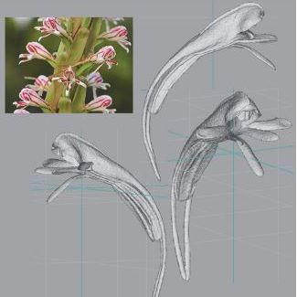
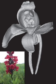
FIG. 1 – 3D reconstruction of the rare Orchid Satyrium Rhodanthum using a Scanco µCT scanner | Due to this, researchers have recently started using innovative micro-CT scanners to obtain 3D reconstructions of low atomic number objects, such as flowers. The great advantage of this method is that it is non-invasive and that it provides extremely fine resolution. Using the Scanco µCT series scanners, studies of geometric morphometrics in plants can be seriously improved. Moreover, pre-scanning treatment of the sample is not required for a fresh-cut flower: the only care remains in correctly positioning the sample to capture its entire section. Since pre-treatment of flowers is unnecessary, this method can be applied also to any flowers that are stored in 70% ethanol. This includes flowers that are collected at remote sites, far away from a micro-CT scanner, and flowers of rare or now extinct species that are stored in herbarium collections. |
Reconstructions of flower surfaces obtained though micro-CT scanning are highly suitable for landmarking processing. This facilitates analysis with images that are visually appealing and easy to interpret, following a relatively simple protocol.

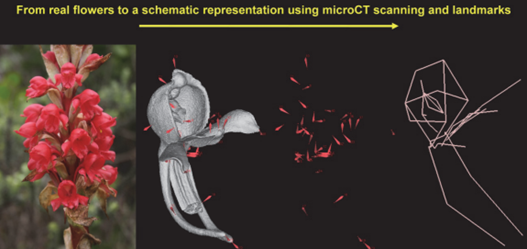
FIG. 2 – Overview of the process to get from real flowers a schematic representation of floral shape using landmarks. Landmarks are shown (without the 3D reconstruction) as red cones on a black background, representing the basic shape of the Orchid flower. If the landmarks relate to lines, a schematic reconstruction of the basic floral shape emerges. This is useful in inter-species comparative analyses.


