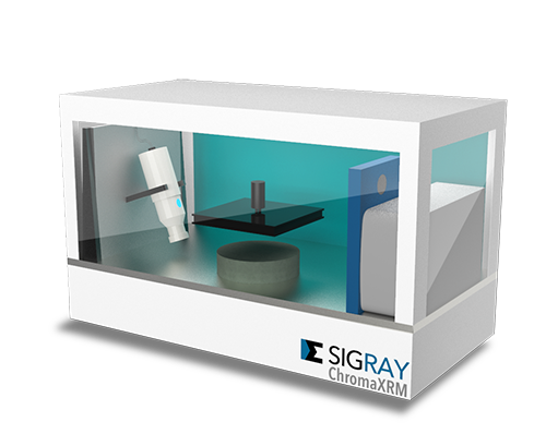
ChromaXRM-500 – Tomographe submicronique pour les sciences de la vie et les polymères
SIGRAYLe nouveau ChromaXRM-500 de Sigray a été spécialement conçu pour répondre aux besoins des applications des sciences de la vie et des polymères, car ces types d'échantillons ne présentent normalement qu'un contraste aux rayons X très limité. Le ChromaXRM-500 dispose d'une nouvelle source de rayons multispectrale brevetée qui peut améliorer le débit d’un facteur 10 grâce à un contraste très élevé.
- Le microscope à rayons X de laboratoire au plus fort contraste d'absorption
- Résolution spatiale submicronique (taille de voxel <300 nm, résolution spatiale de 500 nm)
- Jusqu'à 5 détecteurs dans un seul système
- Encombrement réduit
Plus d'informations
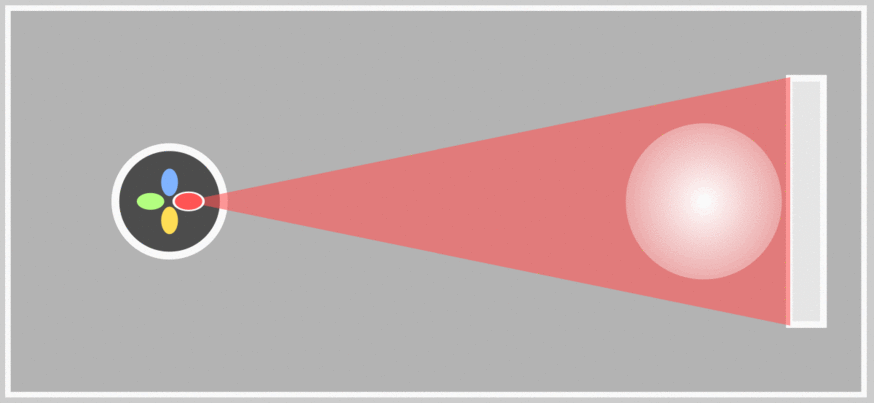
The ChromaXRM-500 is using Sigray´s patented x-ray mulri-color source. The source can be fitted with 5x different targets and offers a very broad energy range and one can use the target that offers the most contrast for the sample’s material leading to a 10x higher throughput.
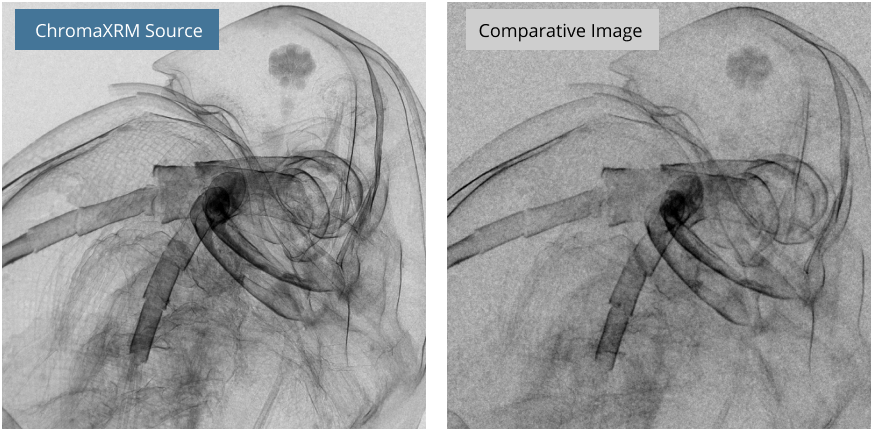
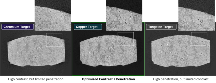
X-rays are generally considered too energetic for Life Science (e.g. soft tissue) or offer only a limited absorption contrast for polymer samples. They are on the other hand sometimes the only choice if you want to get a high-resolution 3D image of your sample without destroying it totally. The ChromaXRM-500 with its 5x user selectable target materials offers now the possibility to optimize the x-ray spectrum for each application and maximize contrast as well as throughput.
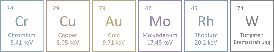
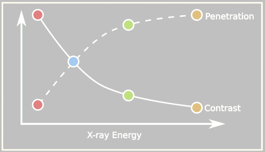
Each target has its own individual energy peak and wavelength comparable to the concept of color in the optical range. The color blue for example has a wavelength of 450nm while red is around 750nm. Comparable to this has a Chromium target its energy peak at 5.41 keV or 2.3 Å while Copper is at 8.05keV or 1.55 Å. Samples of different compositions and sizes are optimized when the illuminating x-ray is of the correct color. Contrast and throughput can be maximized by >10X. Other XRM and microCT products on the market only offer an x-ray source with a Tungsten target, which offers good penetration but poor contrast on many challenging samples.
Caractéristiques techniques
| Parameter | Specification | |
|---|---|---|
| Overall | Spatial Resolution | 0.5 um with 40X objective |
| Minimum Voxel | 275 nm | |
| Source | Type | Sigray patented ultrahigh brightness sealed microfocus source |
| Voltage | 20 - 60 kVp | |
| Power | 100W | |
| Target(s) | Up to 5 targets. Includes selection from Cr, Cu, Rh, W, Mo, Au, Ti, Ag. Others available upon request. | |
| Detector(s) | Type | Up to 5 detectors. Includes LFOV detectors and high resolution detectors. |
| Camera | 4MP deep cooled CCD | |
| Visible Light Camera | 16MP alignment camera | |
| Software | Command and Control | Sigray 3D with Intuitive interface |
| Reconstruction | GigaRecon - fastest commercial CBCT reconstruction software | |
| Offset Scans | Expands the horizontal FOV. Sigray software advantage | |
| Helical Scan | Enabled for tall samples | |
| AutoPilot | AI-assisted microscope operation for unsupervised acquisition | |
| Linux Workstation | Interface is on a Windows workstation, while a separate robust Linux workstation controls the system. Advantageous for reliable 24-7 operation. | |
| EPICS | Open-source software controls for maximum flexibility | |
| Dimensions | Footprint | Desktop system 250 kg |
| Sample Size | 50 x 50 mm diameter |
Applications
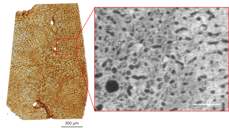
Life Science- unstained liver cells
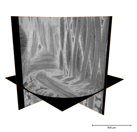
Carbon fibers
Pharmaceuticals
Téléchargements


Contact


