
")

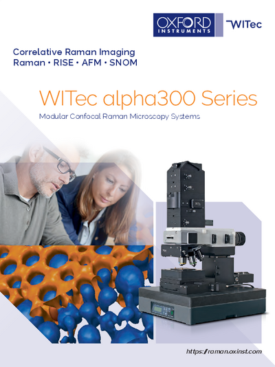
SNOM, confocal microscopy and AFM system
alpha300 S from WITecThe alpha300 S is a user-friendly scanning nearfield optical microscope (SNOM) that combines the advantages of SNOM, confocal microscopy and atomic force microscopy (AFM) in a single instrument. Switching between the different modes can be easily done by rotating the objective turret. The alpha300 S uses unique micro-fabricated SNOM cantilever sensors for optical microscopy with spatial resolution beyond the diffraction limit.
- Spatial resolution beyond the diffraction limit (ca. 60 nm laterally)
- Unique patented SNOM sensor technique
- Ease-of-use in air and liquids
- Various atomic force microscopy (AFM) modes applicable
- Non-destructive imaging technique with minimal, if any, sample preparation
Further information
The WITec scanning near-field optical microscope (SNOM) alpha300 S operates using a unique near-field objective. It is mounted in the objective turret and provides access to SNOM or AFM. The micro-fabricated SNOM sensors are held magnetically at the end of the objective’s arm, enabling simultaneous observation of the cantilever and sample. For quick cantilever alignment, the arm can be moved in all three dimensions by an integrated highly-precise inertial drive. The movements are controlled by the WITec Control software, which also provides convenient alignment routines. The objective not only focuses the excitation laser beam, but also the beam-deflection laser for distance control. Highly focused, ultra-stable optics guarantee low-noise measurements without interference from the two laser systems. Using standard AFM cantilevers, the alpha300 S includes full AFM capability.
Specifications
Scanning near-field optical microscopy (SNOM) modes:
- bottom up and top down
- collection
Confocal microscopy (CM) modes:
- Transmission
- Reflection
- Fluorescence (optional)
SNOM-AFM combinations:
- AFM operation modes of alpha300 A included or optional available
- Acquisition of force-distance curves and light-distance curves
- Fixed-bottom illumination
- Total internal reflection illumination (optional)
Microscope features:
- Research grade optical microscope with 6 x objective turret
- Video system: eyepiece color video camera
- LED white-light source for Köhler illumination of tip and sample
- High sensitivity b/w video camera to view sample and SNOM/AFM tip in transmission
- Manual sample positioning in x- and y-direction, 25 mm travel
- Microscope base with active vibration isolation system
- Piezo-driven scan stage (scan range 100 x 100 x 20 µm; others optional)
Sample size:
- Usually 120 mm in x- and y-direction, 25 mm in height (adapter for larger heights available)
Computer interface:
- WITec software for instrument and measurement control, data evaluation and processing
Applications
Downloads


Contact

Navigation
Categories
Contact
Quantum Design GmbH
Meerstraat 177
B-1852 Grimbergen
Belgium
| Mobil: | +32 495 797175 |
| E-mail: | beneluxqd-europe.com |
