Novel tri-contrast imaging & in-situ optimization with the PrismaXRM 3D x-ray microscope
3D x-ray microscopy (also known as micro-CT) has developed substantially in the past two decades. Many systems are now reaching the limits of resolution achievable at high throughput. PrismaXRM was designed to overcome visibility limits of existing leading x-ray microscopes. The flexible system provides not only the highest spatial resolution on the market, but also a novel, multi-contrast approach that is superior to absorption alone for identifying sub-resolution features, such as cracks and nanoparticles.
PrismaXRM is a novel 3D x-ray microscope that breaks through the visibility limits of the existing leading x-ray microscopes. The breakthrough design combines two independent configurations that may be selected by the user in seconds:
- High submicron resolution absorption micro-CT
- A multi-contrast Talbot-Lau interferometer
The absorption contrast configuration offers capabilities that match and exceed those offered by other premium systems, and the interferometer configuration exclusively adds two contrast mechanisms: Quantitative Phase and Subresolution Darkfield.
What are the advantages of Quantitative Phase and Subresolution Darkfield?
X-ray absorption contrast microscopy has advanced considerably in the past decade to maturity. Many vendors now providing comparable performance of 0.5 to 1 μm spatial resolution, which is near the limit of lensless x-ray microscopy. The PrismaXRM breaks through these barriers with new imaging modalities that overcome the barriers to conventional imaging.
- With Quantitative Phase, the first differential of the refractive index is obtained. This provides not only stronger phase information than edge enhancement phase, but also enables quantitative results for compositional analysis
- With Subresolution Darkfield, features below the resolution (e.g. on the order of 100 s/nm) such as crack tips and voids are seen. Nanoparticle migration and inclusions have also been seen using the PrismaXRM’s Darkfield capabilities
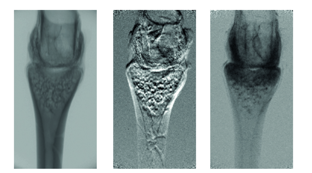
Tri-Contrast Imaging: Sigray PrismaXRM simultaneously acquires three forms of x-ray contrast: absorption, Quantitative Phase, and Subresolution Darkfield. Shown above is a tri-contrast image of a frog’s toe joint using the Sigray PrismaXRM; different features are clearly visible in each mode of contrast, such as the spongey tissue using the darkfield and musculature in phase contrast. Tri-contrast imaging has demonstrated powerful capabilities for a range of new applications such as lithium migration in battery samples, hidden cracks in semiconductor packaging, and more.
Please contact us to receive further information.


