

Inverted Confocal Raman Imaging
witec360 Ri from Oxford InstrumentsThe witec360 Ri turns 3D chemical characterization upside down. Its inverted beam path preserves all the functionality of Oxford Instruments’ standard witec360 confocal Raman imaging microscopes while introducing a new angle in access and handling. The ability to view and investigate samples from below is a great advantage when working with aqueous solutions and oversized samples. Studies in life sciences, biomedicine and geosciences in particular will benefit from the consistency and flexibility provided by the geometry of the witec360 Ri.
- Consistent, repeatable positioning of liquid samples
- Motorized sample stage
- Easy Bulky samples analysis
- Features all imaging and spectroscopy capabilities of the WITec alpha300 R series, included its modularity and expandability
- Non-destructive, contact-free, label-free chemical characterization
- True confocality, ideally suited to 3D image generation
- Lateral resolution limited only by physical law
- Compatible with other microscopy techniques (fluorescence, DIC, phase-contrast, …)
Further information
An inverted beam path allows liquid samples to be placed on the fixed plane of the stage for quick and repeatable measurements.
Bulky samples that would be challenging to investigate underneath a conventional microscope objective turret can be accommodated by placing them on the stage of the witec360 Ri.
The motorized sample stage also facilitates the mounting of environmental enclosures and other accessories.
Application Examples
Correlative Raman-Fluorescence microscopy image of DAPI-marked eukaryotic cell nuclei
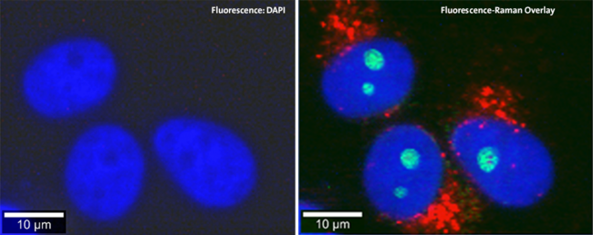
Correlative Raman Fluorescence Microscopy: Fluorescence DAPI Staining & Raman Imaging
3D Raman image of pressed banana pulp
The witec360 Ri combines the advantages of data acquisition from below with the established merits of 3D confocal Raman imaging which is a powerful and versatile technique that can chemically characterize samples nondestructively and without labeling or other specialized preparation.
The image shows a 3D reconstruction of Raman image-stack acquisition of banana pulp.
Scan range: 300µm x 200µm x 90µm
Pixels (Spectra): 450x300x45
Pressed banana pulp sample: Starch grains (green) and cell wall components (red)
Specifications
Raman General Operation Modes
- Raman spectral imaging: Acquisition of complete hyperspectral Raman imaging data sets while continuously scanning.
- Planar (x-y-direction) and depth scans (z-direction) with motorized sample positioning
- Image stacks: 3D confocal Raman imaging
- Fast and slow time series
- Single point Raman spectrum acquisition and depth profiling
- Fibre-coupled Hexalight spectrometer specifically designed for Raman microscopy and applications with low light intensities
- Confocal Fluorescence Microscopy
Basic Microscope Features
- Research grade inverted optical microscope with 6x objective turret
- Video system: Video CCD camera and/or fluorescence camera
- LED white-light source for Köhler illumination
- Binocular
- Condensor lens for up to 7 contrasts (e. g. brightfield, DIC, Phase contrast, NAMC, etc.)
- Sample holder accommodating various standard sample formats (e. g. microscopy slides, Terasakiplates, 35/65 mm dishes, Nunc flasks, counting chambers)
- Internal filter block revolver
- Motorized sample positioning and scanning stage in x- and y-direction, large travel range
Raman Optional/Upgradable Operation modes
- A broad range of lasers and wavelenghts (from UV to IR) eligible
- Highly customizable Hexalight spectrometer (UV, VIS, NIR)
- Ultrafast Raman imaging optional available
- Upgradable for time correlated single photon counting / fluorescence lifetime imaging (FLIM)
- Autofocus
Ultrahigh-throughput Oxford Instruments Hexalight Spectrometers
- Various lens-based, excitation optimized spectrometers (UV, VIS or NIR) available, all specifically designed for Raman microsopy and applications with low light intensities
- Fibre-coupled ultrahigh-throughput optical instruments
- Superior peak shape conservation
Computer Interface:
- Oxford Instruments software for instrument and measurement control, data evaluation and processing
Downloads


Contact


Navigation
Categories
Contact
Quantum Design GmbH
ul. Sztygarska 12/3
41-500 Chorzow
Poland
| Mobile: | +48 515 166893 |
| E-mail: | polandqd-europe.com |
