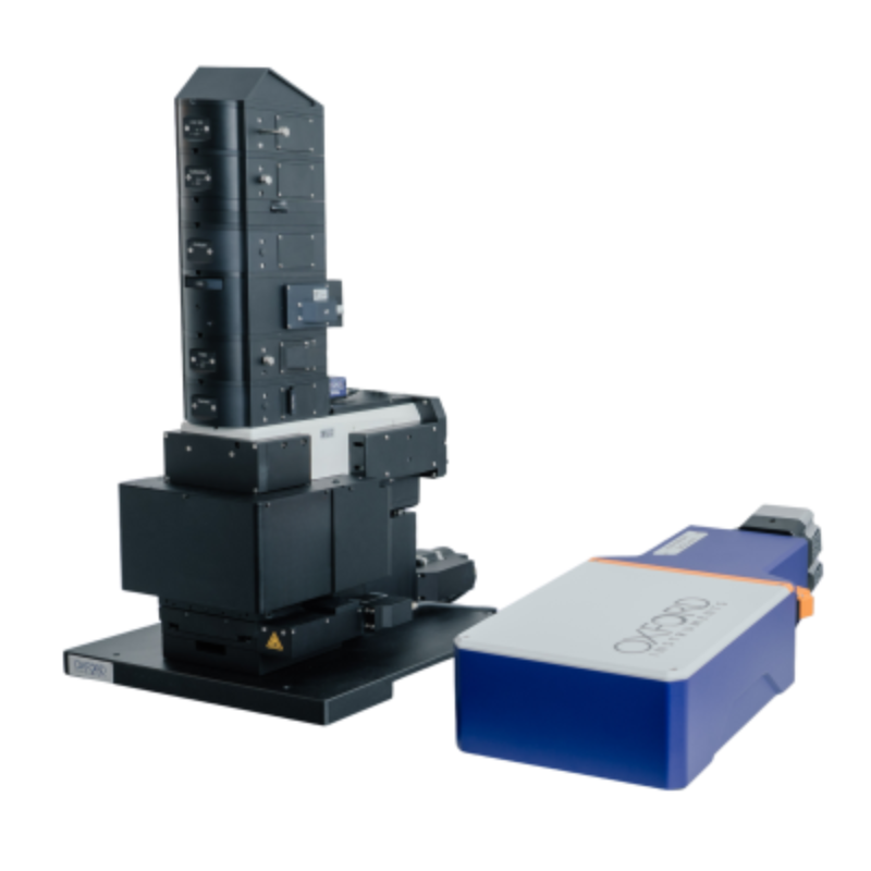


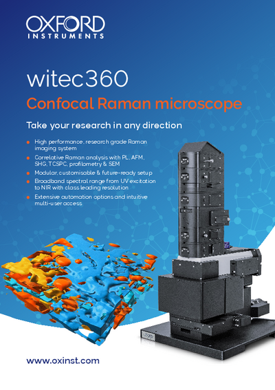
Ultra-high-resolution confocal Raman Microscope
WITEC360 from Oxford IstrumentsThe imaging capabilities of the WITEC360 fulfill the highest requirements of confocal Raman imaging with superior performance in speed, sensitivity and resolution. These unique characteristics have established the WITEC360 the preeminent confocal Raman imaging system on the market.
- Confocal Raman Imaging with unprecedented performance in speed, sensitivity, and resolution
- Hyperspectral image generation with the information of a complete Raman spectrum at every image pixel
- Excellent lateral resolution
- Outstanding depth resolution ideally suited for 3D image generation and depth profiles
- Ultra-fast Raman imaging option with under one millisecond integration time per spectrum
- Ultra-high throughput spectroscopic system for highest sensitivity and best performance in spectral resolution
- Non-destructive imaging technique: no staining of fixation of the sample required
Further information
While long recognized as the state-of-the-art imaging system, ongoing development resulting from Oxford Instruments' innovative spirit has kept the WITEC360 at the forefront of Raman microscopy and set the benchmark in terms of imaging capability as well as spectral quality, spatial resolution, ease-of-use and compatibility with other measurement techniques.
The flexibility of the WITEC360 series allows the system adapt to all requirements, combine different imaging techniques and to evolve to meet new or expanded needs.
The WITEC360 offers the following extensions:
- Ultrafast Raman Imaging (1300 spectra per second) optional available
- Wide choice of lasers and possibility to install additional lasers
- Dark Field, Phase Contrast and DIC optional
- Upgradable for epi-fluorescence applications
- Piezo-driven scan stage (scan range 200 x 200 x 20 µm) (other ranges available)
- TrueSurface for Raman depth profiling
Specifications
Raman General Operation Modes:
- Raman spectral imaging: acquisition of a complete Raman spectra at every image pixel
- Planar (x-y-direction) and depth scans (z-direction) with manual sample positioning
- Image stacks: 3D confocal Raman Imaging
- Time series
- Single point Raman spectrum acquisition
- Single-point depth profiling
- Fibre-coupled Hexalight spectrometer specifically designed for Raman microsopy and applications with low light intensities
- Confocal fluorescence microscopy
- Bright Field Microscopy
Basic Microscope Features:
- Research grade optical microscope with 6x objective turret
- Video system: video CCD camera
- LED white-light source for Köhler illumination
- Manual sample positioning in x- and y-direction, optional high-resolution motorized stage with various travel ranges
- Fibre coupling
Raman Optional/Upgradable Operation modes
- Additional lasers, several wavelenghts eligible
- Extensive versatility and configurability of the Hexalight spectrometer (UV, VIS, NIR)
- Automated, motorized sample positioning and measuring with piezo-driven scan stages
- Automated confocal Raman imaging
- Automated multi-area and multi-point measurements
- Full automation capability
- Ultrafast Raman imaging, optional available
- Upgradable for epi-fluorescence applications
- Adapter for higher samples
- TrueSurface for Raman depth profiling
- Autofocus
- Dark Field Microscopy, Phase Contrast Microscopy, and DIC optional
Features of the Hexalight Ultrahigh-Throughput Spectrometer
- Various lens-based, excitation optimized spectrometers (UV, VIS or NIR) available, all specifically designed for Raman microsopy and applications with low light intensities
- Fibre-coupled ultrahigh-throughput optical instruments
- Superior peak shape conservation
Computer Interface:
- Oxford Instruments software for instrument and measurement control, data evaluation and processing
Applications
From measurements in liquids to solid samples or soft tissues in life science varied samples are analyzed on a regular basis. With their convenient handling and versatile analytical capabilities the flexible WITec imaging systems provide the opportunity to adjust the imaging technique to changing requirements and are particularly well-suited for life science.
The development and production of drug delivery systems requires efficient and reliable control mechanisms to ensure the quality of the final products. These products can vary widely in composition and application. Therefore analytical tools such as the WITec imaging systems that provide both comprehensive chemical characterization and the flexibility to adjust the method to the investigated specimen are preferred in pharmaceutical research.
Materials science is a diverse field including the development and testing of new substances, as well as the refinement of manufacturing processes and quality control for existing products. WITec imaging systems are particularly well-suited for comprehensive sample analyses in materials science and provide the opportunity to acquire a thorough knowledge of the sample surface morphology and chemical composition.
WITec confocal Raman imaging systems are excellent analytical tools for the comprehensive investigation of geological samples, such as the identification and characterization of minerals, or in the observation of mineral phase transitions in high and ultra-high pressure/temperature experiments.
WITec imaging systems enable comprehensive sample analysis that provides a thorough characterization of the physical and chemical properties of the polymers on the nanometer scale.
2D materials such as carbon nano-tubes, graphene or transition metal dichalcogenides (TMDs) show immense promise in many applications such as transistors, sensors, and optoelectronics. Flexible and adaptive analytical methods can support effective investigation and accelerate progress in 2D materials research and development. For comprehensive investigations of nano-carbon and TMD samples WITec microscopes can be equipped with various imaging techniques such as confocal Raman imaging, AFM, and SNOM, all fully integrable in a single microscope.
Downloads


Contact


Navigation
Categories
Contact
Quantum Design s.r.l.
Via di Grotta Perfetta, 643
00142 Roma
Italy
| Phone: | +39 06 5004204 |
| Fax: | +39 06 5010389 |
| E-mail: | italy@qd-europe.com |
