


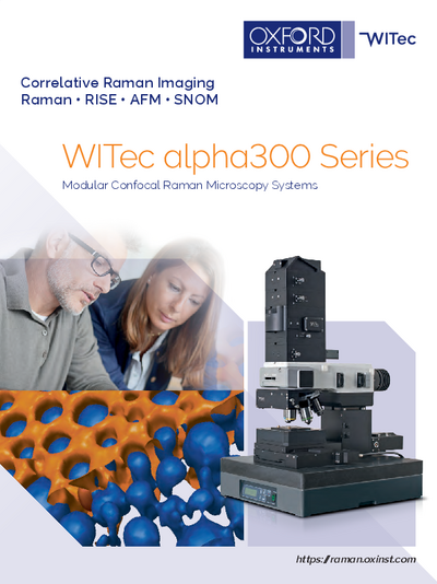
Our partner

Combined Raman and scanning nearfield optical microscopy (SNOM) system
alpha300 RS from WITecFor the user with challenging experimental requirements, the alpha300 RS facilitates confocal Raman imaging in combination with scanning near-field optical microscopy (SNOM) for optical imaging with resolution beyond the diffraction limit. It combines all the features of the alpha300 S and alpha300 R and many AFM operation modes. Furthermore the combined Raman-SNOM microscope is ideally suited for combined high-resolution Raman imaging techniques such as nearfield-Raman imaging.
- Provides all features of the alpha300 R (Raman) and the alpha300 S (SNOM) microscope in one instrument
- Excellent combination of high-resolution surface imaging (SNOM) and chemical imaging (Raman)
- Ideally suited for combined techniques such as nearfield-Raman imaging
- Switch between the measurement techniques is realized by a rotation of the objective turret
- No sample movement between the measurements
Further information
The alpha 300RS offers the following extensions:
- alpha300 RS+ for automated measurements
- Automated motorized sample positioner in x-, y-, and z-direction, 25 mm travel range (50 mm travel range optional)
- Automated confocal Raman Imaging (25 x 25 mm; optional 50 x 50 mm)
- Automated multi-area and multi-point measurements
- 2D and 3D Raman mapping
- Raman depth profiling
- Autofocus
Specifications
Raman operation modes:
- Raman spectral imaging: acquisition of a complete Raman spectra at every image pixel
- Planar (x-y-direction) and depth scans (z-direction)
- Image stacks: 3D confocal Raman Imaging
- Time series
- Single point Raman spectrum acquisition
- Single-point depth profiling
- Ultrafast Raman Imaging (1300 spectra per second) optional available
- Confocal fluorescence microscopy
- Bright Field Microscopy
- Dark Field, Phase Contrast and DIC optional
- Upgradable for epi-fluorescence applications
- Dark Field, Phase Contrast and DIC optional
- SNOM Operation Modes
- Scanning Near-field Optical Microscopy (SNOM) modes: bottom up and top down mode, collection mode
Confocal microscopy (CM) modes:
- Transmission
- Reflection
- Fluorescence (optional)
- SNOM-AFM combinations: AFM operation modes of alpha300 A included or optional available
- Acquisition of force-distance curves and light-distance curves
- Fixed-bottom illumination
- Total internal reflection illumination (optional)
- AFM Operation Modes:
- AFM Modes
- Contact Mode
- Lateral Force Mode
Microscope features:
- Research grade optical microscope with 6 x objective turret
- Video system: eyepiece color video camera
- LED white-light source for Köhler illumination of tip and sample
- High sensitivity b/w video camera to view sample and SNOM/AFM tip in transmission
- Manual sample positioning in x- and y-direction, 25 mm travel
- Microscope base with active vibration isolation system
- Piezo-driven scan stage (scan range 100 x 100 x 20 µm; others optional)
Sample size:
- Usually 120 mm in x- and y-direction, 25 mm in height (adapter for larger heights available)
Computer interface:
- WITec software for instrument and measurement control, data evaluation and processing
Applications
Life science
From measurements in liquids to solid samples or soft tissues in life science varied samples are analyzed on a regular basis. With their convenient handling and versatile analytical capabilities the flexible WITec imaging systems provide the opportunity to adjust the imaging technique to changing requirements and are particularly well-suited for life science.
Pharmaceutics, cosmetics
The development and production of drug delivery systems requires efficient and reliable control mechanisms to ensure the quality of the final products. These products can vary widely in composition and application. Therefore analytical tools such as the WITec imaging systems that provide both comprehensive chemical characterization and the flexibility to adjust the method to the investigated specimen are preferred in pharmaceutical research.
Materials science
Materials science is a diverse field including the development and testing of new substances, as well as the refinement of manufacturing processes and quality control for existing products. WITec imaging systems are particularly well-suited for comprehensive sample analyses in materials science and provide the opportunity to acquire a thorough knowledge of the sample surface morphology and chemical composition.
Geoscience
WITec confocal Raman imaging systems are excellent analytical tools for the comprehensive investigation of geological samples, such as the identification and characterization of minerals, or in the observation of mineral phase transitions in high and ultra-high pressure/temperature experiments.
Polymers
WITec imaging systems enable comprehensive sample analysis that provides a thorough characterization of the physical and chemical properties of the polymers on the nanometer scale.
Nano-Carbon and graphene
Nano-carbon materials such as graphene or carbon nano-tubes show immense promise in many applications such as transistors, sensors, and optoelectronics. Flexible and adaptive analytical methods can support effective investigation and accelerate progress in nano-carbon & graphene research and development.
Downloads

Contact

Dr.
Agnieszka
Kowalczyk-Głowacka
Navigation
Categories
Contact
Quantum Design GmbH
ul. Sztygarska 12/3
41-500 Chorzow
Poland
| Phone: | +48 32 2482048 |
| Mobile: | +48 515 166893 |
| E-mail: | polandqd-europe.com |
