Magnetic Force Microscopy (MFM)
The magnetic force microscopy scanning technique, or MFM, described here is the main method of scanning probe microscopy to probe samples with magnetic properties or magnetic materials and elucidate features such as magnetic domains and domain walls in a sample. A typica application is in the field of magnetic storage media for quality control.
In MFM, the magnetic forces acting on a sharp, magnetized tip by the sample are measured. During this measurement the tip is lifted off the surface to separate the long-range magnetic forces from the short-range atomic forces between tip and sample. Magnetic force microscopy operates in amplitude modulation dynamic mode, this mode is also referred to as tapping mode. The cantilevers are inexpensive and commercially available. MFM maps the phase and frequency of the oscillating cantilever as it passes over at a prescribed height over the sample. A repulsive magnetic force gradient will cause the resonance curve to shift to a higher frequency, accompanied by an increase in phase shift (bright contrast). Conversely, an attractive magnetic force gradient results in the resonance curve shifting to a lower frequency, accompanied by a decrease in phase shift (dark contrast). The advantages of MFM operation in dynamic mode are lower noise and higher resolution. The tip-sample distance is a crucial parameter to optimize for effective MFM operation. If the tip is too far from the sample, the resolution will be compromised. If the tip is too close to the sample, the topography will be convoluted into the MFM signal, significantly complicating its interpretation.
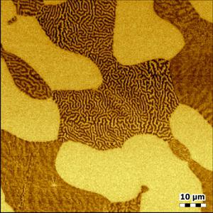
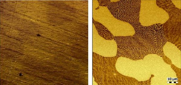
Below are MFM images of polished stainless steel. On the left is an 80µm x 80µm topography image showing a very smooth surface with some polishing marks running diagonally across the surface. The right image is the corresponding MFM phase image for this area, revealing very distinct features and morphology not evident in the topography image. The MFM clearly shows magnetic domains and the magnetic ferrite phase in the maze-like pattern.
Magnetic Force Microscopy in applied magnetic fields
The Nanosurf AFM suite can be equipped with a variable magnetic field sample holder which allows to apply a DC magnetic field in-plane with the sample. MFM imaging can be performed with the sample in a DC magnetic field up to 720mT. Areas of application are magnetic imaging of ferromagnetic films and nanostructures. As an example, below are a series of MFM images 7µm x 7µm of Shakti lattice under different applied magnetic fields (from left to right, -20mT, -50mT and -200mT).

Discover our AFM range here >>
| +39 06 5004204 | |
| +39 06 5010389 | |
| pergoliniqd-europe.com |

