Cryo preparation system for scanning electron microscopy
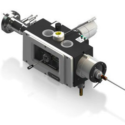
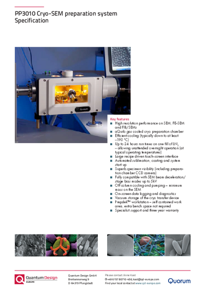
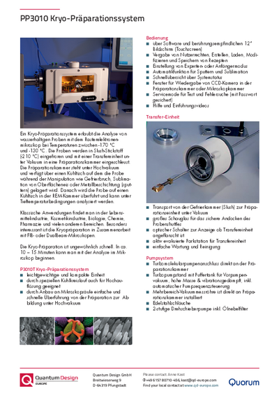
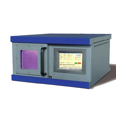
PP3010 from Quorum TechnologyThe cryo preparation system allows the analysis of wet samples with scanning electron microscopy (SEM) at temperatures down to -190 °C. During cryo preparation, samples are frozen rapidly and fed under vacuum conditions to a preparation chamber by means of a transfer unit. The preparation chamber is under high vacuum and fitted with a cold stage on which the sample rests during manipulations like cold fracturing, sublimation of surface ice or metal / carbon coating. The sample is then transferred to a cold stage in the SEM chamber for further analysis under cryogenic conditions.
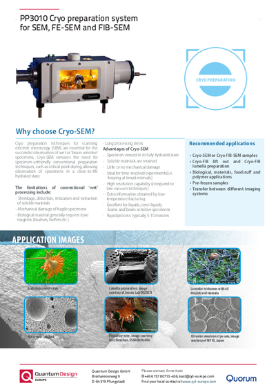
Cryo preparation is exceptionally fast - microscopic analysis can start in only 10 – 15 minutes. Typical applications can be found in the food and cosmetics industry, biology, chemistry, pharmacy, and other sectors. Cryo preparation is particularly interesting in conjunction with FIB or DualBeam microscopes.
- Gas cooled cryo preparation chamber
- Efficient cooling down to -190 °C
- 24 hours plus run times on one fill of LN2
- Easy operation via touch screen
- Automatic function for sputtering and sublimation
Further information
The PP3010 cryo preparation system is column-mounted. This means the sample does not have to be returned to the lower-vacuum transfer unit for further optimization of the sample surface. Further optimization may include water sublimation, cold fracturing at a different point, and subsequent coating with precious metal or carbon.
The system is software-controlled, which allows the automation of many of the working steps. Recipes and system configurations can be created, modified, loaded and stored. The award of user rights allows the system to be used by several users which increases sample throughput.
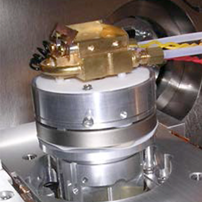
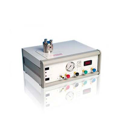
PP3010 uses N2 gas for cooling. The gas is cooled by liquid nitrogen through heat exchangers. Cooling with nitrogen gas is absolutely free of vibration. All cooled units (cold trap, cold stages in the preparation and microscope chamber) are independently cooled and controlled. The intelligent design ensures that the cold trap always remains the coldest part in the preparation chamber, and offers an efficient temperature gradient relative to both cold stage and sample. It can be freely positioned within the microscope chamber, even between pole shoe and sample, if necessary.
Three ceramic balls ensure low-temperature decoupling between stage and sample holder. Ceramics are insulators and do not change their volume under the influence of alternating temperatures. The sample holder’s dovetail adapters in both the preparation and microscope chambers use a single clip and are optimized for easy specimen introduction.
Both the preparation and SEM chambers are fitted with a CCD camera. The images are displayed on the operator screen which facilitates the loading of the cold stage into the SEM with the transfer unit. The process can be observed through several viewing windows and by the CCD camera image displayed on the touch screen. LED lamps in the preparation unit and the microscope chamber generate cold light and facilitate visual inspection.
The preparation system is equipped with a steel blade with active cooling for cold fracturing. Cold fracturing always takes place at the points with the lowest resistance or the points with the lowest bond forces. Height can be adjusted with the help of a micrometer gauge attached to the blade. However, this merely adjusts and monitors the point at which the blade is applied for inducing the cold fracture (z-axis), not where the sample will actually break. It cannot be used as substitute microtome.
For high resolution applications, platinum is frequently used as a coating material. Sputtering requires argon which cannot be cooled down to liquid N2 temperatures because it will freeze. The more and the longer argon is used, the more the temperature in the preparation chamber rises – which is an absolutely undesirable effect. The PP3010T system feeds argon gas directly into the sophisticated magnetron sputter head.
Thanks to the elaborate configuration of the head, plasma remains concentrated underneath the target to the largest possible extent. Normally, the applied voltage accelerates free electrons towards the sample and the kinetic energy that is generated when the electrons hit the sample cause the sample to heat. This effect should be avoided or at least reduced to a minimum. With PP3010, electrons which are generated during ionisation are directed away from the sample. With a Pt target, a magnification of 100,000 x can be achieved easily.
Specifications
| Inside microscope | |
|---|---|
| Gas cooled cold stage | with integrated heaters and temperature sensor |
| Gas cooled anti-contaminator (cold trap) and variable position mount | including temperature sensor. Shape, size and mounting position tailored to the microscope |
| Cold stage and anti-contaminator (cold trap) | capable of reaching -190 °C or lower within 10 minutes of cool down start. Temperature stability of <1 °C. Maximum cold stage temp >100 °C |
| CCD camera | with image displayed on main control screen (only available if the microscope geometry allows) |
| Cold stage parking bracket | for when the system is not in use (only fitted if the microscope chamber geometry allows) |
| Cooling system for SEM cold stage and anticontaminator, and cryo preparation chamber cold stage and cryo shields | |
| CHE3010 off-column | 21 l nitrogen dewar and heat exchanger. For cooling both cold stages and all cold traps, typically with up to 24 hour hold times at normal operating temperatures |
| Independent circuits | for SEM cold stage and SEM anti-contaminator |
| Electronic gas flow control | valves with flow sensors |
| On the microscope column | |
| Feed-through | for cold gas supply to the cold stage |
| Suitable adapter flange | for the cryo preparation chamber |
| aQuilo cryo preparation chamber | |
| aQuilo cryo preparation chamber | mounted directly to the SEM using a suitable microscope chamber port |
| Integrated gate valves | with solenoid interlock to prevent accidental opening |
| Nitrogen gas cooled cold stage | with heater and temperature sensor integrated into the cold stage to give variable temperature control from > +100 °C to less than -190 °C |
| Nitrogen gas cooled cold traps around specimen | for trapping water vapour. Temperature: close to liquid nitrogen for maximum cold trapping capabilities |
| High specimen visibility | Large front window (150mm x 78mm) plus two top viewing ports |
| CCD camera | to observe specimen fracturing and pickup |
| Cold fracturing | The cryo preparation chamber is fitted as standard with a front mounted fracturing and manipulation device. The ball-jointed mount allows flexible movement of the blade. The device is fitted with a no.5 scalpel (interchangeable with other scalpel blades – not supplied) |
| Micrometer advanced rigid bladed, fracturing tool | available as an option |
| Fully integrated, automatic, high resolution sputtering system | including a platinum (Pt) target (E7400-314C). Other targets, including, Au, Au/Pd, Cr and Ir are available as options |
| Multi-range vacuum gauge | fitted directly to the chamber |
| Chamber lighting | three bright LED lamps focused on the specimen stage |
| Opto-switch sensing of cryo transfer device | being fitted to the chamber airlock |
| Airlock control switches and LEDs | located on cryo preparation chamber |
| Pumping system | |
| Stand alone turbo stack | 70 l/s |
| Base vacuum | 1-6 mbar |
| Stainless steel bellows | connection to preparation chamber |
| Rrotary vacuum pump | 5 m3/h (required – order separately) |
Applications
Typical applications can be found in the food and cosmetics industry, biology, chemistry, pharmacy, and other sectors. Cryo preparation is particularly interesting in conjunction with FIB or DualBeam microscopes.
Downloads
Videos
Contact
| +49 6157 80710-12 | |
| +49 6157 80710912 | |
| Write e-mail |

| +49 6157 80710-456 | |
| +49 6157 807109456 | |
| Write e-mail |


